dental x ray positioning guide
Dental X-ray positioning is crucial for accurate imaging, ensuring clear visualization of dental structures. Proper techniques minimize radiation exposure and enhance diagnostic accuracy, essential for effective treatment planning.
Overview of Dental Radiography
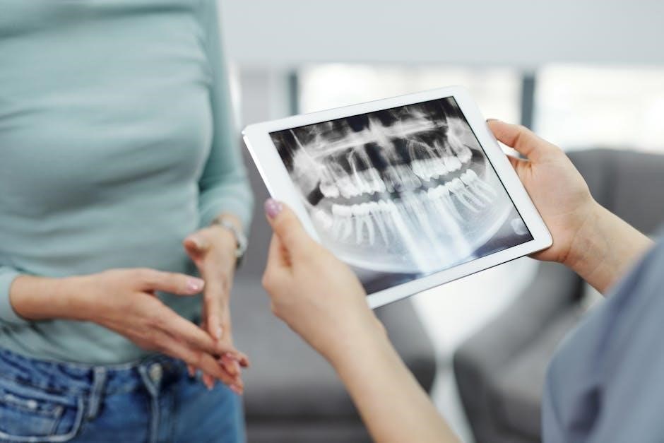
Dental radiography is a fundamental diagnostic tool in dentistry, utilizing X-rays to capture detailed images of teeth, gums, and surrounding structures. It plays a vital role in detecting cavities, abscesses, and bone loss. Intraoral X-rays, such as periapical and bitewing views, provide precise details of individual teeth and their supporting tissues. Extraoral X-rays, like panoramic images, offer a broader view of the jaw and facial bones. Proper positioning is essential to ensure clear, accurate images while minimizing radiation exposure. Advances in digital radiography have enhanced image quality and reduced radiation doses, making the process safer and more efficient for both patients and clinicians. Regular X-rays are recommended for maintaining optimal oral health and early detection of potential issues.
Importance of Proper Positioning in Dental X-Rays

Proper positioning in dental X-rays is critical for obtaining clear, accurate images essential for diagnosis. Misalignment can lead to distorted images, potentially causing misdiagnoses or unnecessary retakes. Correct positioning ensures that all relevant structures are captured, reducing the need for additional imaging and minimizing radiation exposure. It also enhances patient safety by allowing clinicians to identify issues early, such as decay or fractures, and plan appropriate treatments. Properly positioned X-rays improve efficiency in dental workflows and provide reliable data for patient care. By adhering to established positioning guidelines, dental professionals can ensure high-quality radiographs that support accurate and effective treatment outcomes, ultimately benefiting patient health and satisfaction.

Types of Dental X-Ray Projections
Dental X-ray projections include bitewing, periapical, and panoramic views. Each type targets specific areas, ensuring comprehensive imaging for accurate diagnosis and treatment planning in dental care.
Bitewing X-Rays: Purpose and Positioning
Bitewing X-rays are essential for detecting interdental cavities and assessing bone levels around teeth; The patient bites a wing-shaped tab to position the film, ensuring accurate imaging. Proper angulation is critical to avoid distortion. This technique minimizes radiation exposure and provides clear views of areas invisible during a routine exam. Regular bitewing X-rays aid in early detection of dental issues, preventing severe problems. They are particularly useful for patients at higher risk of caries or periodontal disease. Correct positioning involves aligning the X-ray beam perpendicular to the film, ensuring optimal results. This method is vital for diagnosing and monitoring dental health effectively.
Periapical X-Rays: Techniques and Relevance
Periapical X-rays provide detailed images of entire teeth, including roots and surrounding bone. Proper positioning involves placing the film parallel to the dental arch, with the X-ray beam angled at 90 degrees. This technique ensures minimal distortion, offering clear views of apical regions. It is invaluable for diagnosing conditions like abscesses, fractures, and periodontal disease. The relevance of periapical X-rays lies in their ability to reveal structural abnormalities not visible in other projections. By capturing the full anatomy of teeth and their supporting structures, they aid in precise treatment planning. Proper alignment and angulation are critical to obtaining diagnostic-quality images, making this projection a cornerstone in dental radiography.
Panoramic X-Rays: Wide-View Imaging
Panoramic X-rays provide a broad overview of the dental and facial structures, capturing the entire maxillofacial region in a single image. This technique involves rotating the X-ray beam around the patient’s head, creating a wide-field view. Proper positioning requires the patient to stand upright with the midline of the face aligned with the beam. The resulting image includes teeth, jaws, sinuses, and the temporomandibular joints (TMJ). Panoramic X-rays are particularly useful for assessing overall dental health, detecting impacted teeth, and evaluating jaw fractures or abnormalities. Their wide coverage makes them ideal for treatment planning in orthodontics, implants, and surgical cases. While they offer less detail than intraoral X-rays, their comprehensive view is invaluable for diagnostic purposes.


Patient Preparation for Dental X-Rays
Patient preparation involves removing jewelry, wearing appropriate clothing, ensuring proper positioning, and following specific guidelines for safety and accurate results.
Positioning the Patient for Optimal Results
Proper patient positioning is essential for obtaining clear and accurate dental X-ray images. The patient should be seated upright or lying down, depending on the type of X-ray, ensuring the head is stabilized. The X-ray beam must be aligned parallel to the dental film or sensor, with correct angulation to avoid distortion. Proper alignment ensures that the resulting images are free from overlap and provide a true representation of the dental structures. Using positioning guides and tools helps maintain consistency and accuracy. Improper positioning can lead to retakes, increasing radiation exposure and delaying diagnosis. By following established protocols, dental professionals can achieve high-quality images while minimizing risks. This step is critical for effective treatment planning and patient safety.
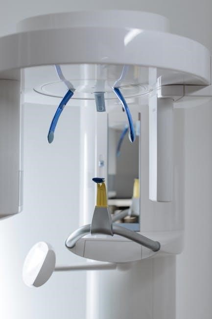
Using X-Ray Positioning Guides and Tools
X-ray positioning guides and tools are indispensable for achieving precise and consistent results in dental radiography. These tools help standardize the alignment of the X-ray beam, ensuring accurate positioning of the patient and the sensor. Positioning guides minimize errors and retakes, reducing radiation exposure and improving image quality. They are designed to accommodate various dental projections, such as bitewing and periapical X-rays. Tools like the iM3 Complete X-Ray Positioning Kit provide clear markers for proper angulation and alignment. By using these guides, dental professionals can ensure that images are captured efficiently and effectively, facilitating accurate diagnoses. Proper use of positioning tools is a cornerstone of modern dental imaging, enhancing both safety and diagnostic outcomes.
Advanced Positioning Techniques
Advanced techniques like digital radiography and cone-beam CT optimize imaging accuracy, reducing radiation exposure and enhancing diagnostic precision with precise patient positioning and cutting-edge technology integration.
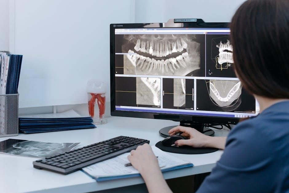
Digital Radiography and Its Impact on Positioning
Digital radiography revolutionizes dental imaging by using electronic sensors and specialized software to capture high-quality images. This technology reduces radiation exposure and processing time, enhancing patient safety. Advanced tools like the iM3 X-Ray Positioning Kit streamline the process, ensuring precise angulation and alignment. Digital systems allow for immediate image preview, enabling quick adjustments for optimal results. Real-time adjustments improve diagnostic accuracy, reducing retakes. Integrated software offers enhanced image clarity and measurement tools, aiding in detailed analysis. This shift from analog to digital methods has transformed dental radiography, emphasizing efficiency and precision. The ability to store and share images digitally further supports collaborative care. Overall, digital radiography positions itself as a cornerstone of modern dental imaging, balancing accuracy, safety, and convenience for both clinicians and patients.
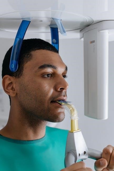
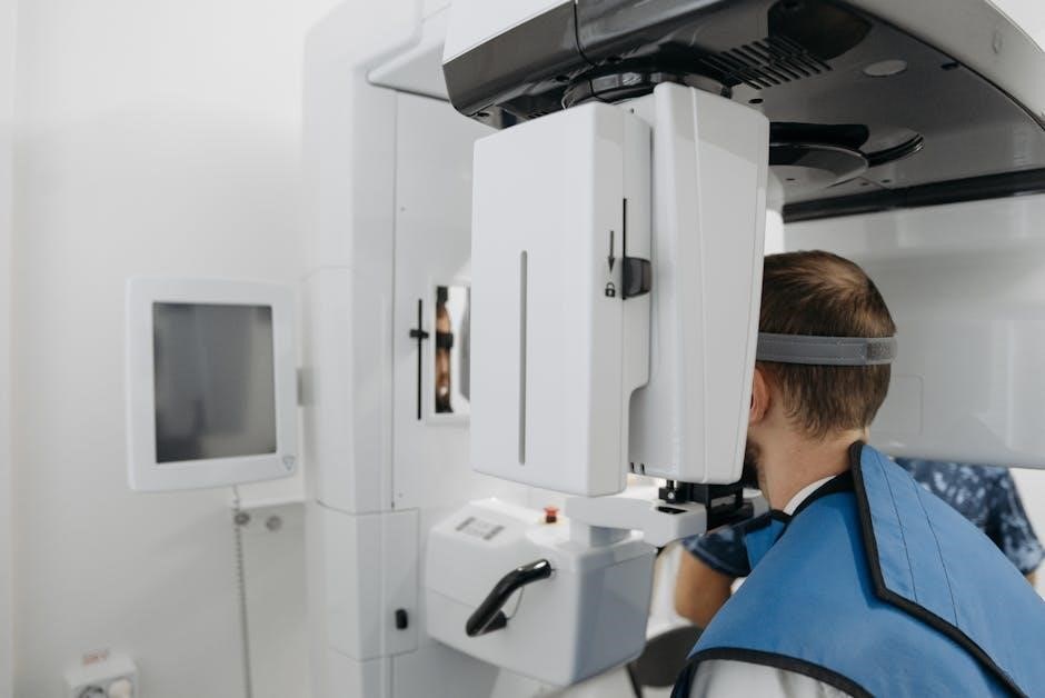
3D Imaging and Cone-Beam CT (CBCT) Positioning
Cone-Beam CT (CBCT) positioning is a advanced technique in dental imaging, offering 3D visualization of dental and facial structures. Unlike traditional X-rays, CBCT provides detailed, high-resolution images, enabling precise diagnosis and treatment planning. Proper positioning is critical to ensure accurate data capture and minimize distortion. The patient is typically seated or standing upright, with the head stabilized to maintain alignment. CBCT systems use rotational scans, capturing data from multiple angles. This method reduces overlapping structures and enhances clarity. Positioning guides and tools, such as headrests and bite blocks, help maintain consistency. CBCT is particularly valuable for complex cases, including implants, orthodontics, and maxillofacial surgery. Its ability to provide a comprehensive view makes it indispensable for modern dental care, improving outcomes and reducing the need for retakes.
Common Mistakes in Dental X-Ray Positioning
Common errors include improper alignment, overexposure, and failure to use positioning guides, leading to distorted images and increased radiation exposure. Avoiding these ensures accurate and safe diagnostics.
How to Avoid Errors in Radiographic Techniques
To avoid errors in radiographic techniques, proper training and adherence to established protocols are essential. Use positioning guides to ensure correct alignment of the X-ray beam and patient anatomy. Regular equipment maintenance and calibration can prevent technical issues. Patient preparation, such as removing metal objects and ensuring proper stabilization, is crucial. Minimize movement by using bite blocks or stabilizing devices. Overexposure can be avoided by adjusting settings based on patient size and anatomy. Consistent monitoring and feedback from experienced professionals help refine techniques. By following these steps, dental professionals can produce high-quality radiographs while minimizing errors and ensuring patient safety.

Best Practices for Dental X-Ray Safety
Adhering to radiation safety protocols is vital in dental imaging. Use lead aprons, thyroid collars, and digital sensors to minimize exposure. Follow ALARA principles to optimize safety.
Radiation Safety Measures in Dental Imaging
Radiation safety is paramount in dental imaging to protect patients and staff from unnecessary exposure. Use lead aprons, thyroid collars, and digital sensors to minimize radiation absorption. The ALARA principle (As Low As Reasonably Achievable) guides dose reduction. Ensure proper equipment calibration and regular maintenance to optimize safety. Digital radiography reduces radiation exposure compared to traditional film. Patient positioning and beam alignment are critical to avoid retakes; Limit X-ray use to diagnostic necessity and inform patients about risks and benefits. Strict adherence to safety protocols ensures compliance with regulatory standards while maintaining image quality for accurate diagnoses.


Leave a Reply
You must be logged in to post a comment.